
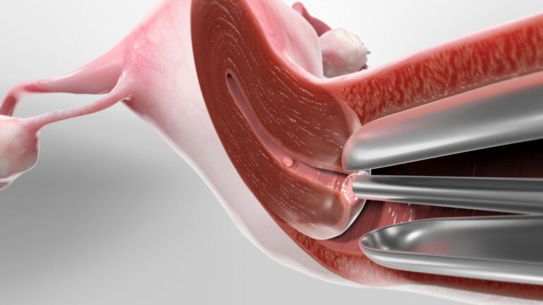
Hysteroscopy is a minimally invasive procedure used to examine and treat issues within the uterus. It involves inserting a hysteroscope, a thin, lighted tube equipped with a camera, through the vagina and cervix into the uterus. This allows the doctor to view the inside of the uterus on a monitor and perform various diagnostic and therapeutic procedures such as removing polyps, fibroids, or intrauterine adhesions, evaluating abnormal bleeding, investigating fertility issues, and collecting biopsies for further examination.
Operative Laparoscopy & Hysteroscopy
Data Research
Excepteur sint occaecat cupidatat non proident.
Data Product
Excepteur sint occaecat cupidatat non proident.
Data Security
Excepteur sint occaecat cupidatat non proident.
Successful Projects
Curabitur in eleifend turpis, id vehicula odio soluta nobis.
Creative Approach
Curabitur in eleifend turpis, id vehicula odio soluta nobis.
How Hysteroscopy Works
Preparation
The patient is typically given a local, regional, or general anesthetic, depending on the complexity of the procedure and the patient’s comfort.
Dilation
The cervix is gently dilated to allow the hysteroscope to pass through.
Insertion of Hysteroscope
The hysteroscope is inserted through the vagina and cervix into the uterine cavity.
Visualization
Saline or carbon dioxide gas may be introduced to expand the uterus, providing a clearer view of the uterine lining.
Examination and Treatment
The doctor examines the uterus for any abnormalities. If necessary, small instruments can be inserted through the hysteroscope to perform procedures such as biopsy, polyp removal, or fibroid resection.
Completion
After the examination and any necessary treatments, the hysteroscope is removed.
Types of Hysteroscopy
Purpose
To diagnose uterine conditions by directly visualizing the uterine cavity.
Uses
Investigating abnormal uterine bleeding, recurrent miscarriages, infertility, or to confirm the presence of uterine abnormalities such as polyps, fibroids, or adhesions.
Purpose
To treat diagnosed conditions within the uterus.
Uses
Removing polyps or fibroids, cutting adhesions (Asherman’s syndrome), removing a uterine septum, or performing endometrial ablation (destruction of the uterine lining to treat heavy menstrual bleeding).
Benefits of Hysteroscopy
Minimally Invasive:
No large incisions are needed, reducing recovery time and discomfort. Typically performed on an outpatient basis, allowing patients to go home the same day.
Direct Visualization:
Provides a clear and direct view of the uterine cavity, allowing accurate diagnosis and treatment.
Combination of Diagnosis and Treatment
Allows for both diagnosis and treatment in a single procedure, reducing the need for multiple surgeries.
Fewer Complications
Lower risk of infection and other complications compared to more invasive surgical techniques.
Risks and Limitations
Risks
Infection
There is a slight risk of developing an infection after the procedure.
Bleeding
Minor bleeding or spotting is common, but significant bleeding is rare.
Uterine Perforation
Accidental puncturing of the uterine wall, though rare, can occur.
Fluid Imbalance
If saline or gas is used to expand the uterus, there is a risk of fluid overload or imbalance.
Limitations
Accessibility
May not be suitable for all patients, especially those with severe cervical stenosis or certain uterine anomalies.
Not a Replacement for All Procedures
Some uterine conditions may still require more invasive surgical techniques.
Preparation and Recovery
Preparation
Medical Evaluation
Preoperative assessment including medical history and any necessary diagnostic tests.
Fasting
Patients may be asked to fast for a certain period before the procedure, especially if general anesthesia is used.
Medication
Instructions on which medications to take or avoid before the procedure.
Recovery
Postoperative Care
Patients are monitored for a short period after the procedure. If local anesthesia is used, recovery time is minimal.
Pain Management
Mild cramping and discomfort are common and can usually be managed with over-the-counter pain relievers.
Activity Restrictions
Patients are typically advised to avoid heavy lifting, strenuous activity, and sexual intercourse for a few days to a week.
Follow-Up
A follow-up appointment may be scheduled to discuss the results and ensure proper healing.